Arteries In Neck Labeled - Practical 2 - Anatomy & Physiology 2102 with Prof.harman ... - Labeled diagram of the arteries of the head and neck.. We go into great detail on the flow of. Ninja nerds!join us in this video where we discuss the blood circulation of the head and neck using a flow chart. The carotids reside beneath the skin on either side, and the pulse can be felt easily with your hand. Cholesterol plaque may slowly build up in the carotid artery wall, over decades. Label the arteries of the neck in the ct angiogram.
The peripheral nervous system is extremely complex as regards the otolaryngologic anatomy and was also segmented in several. Cholesterol plaque may slowly build up in the carotid artery wall, over decades. It is located on every side of the neck and is a large triangular space, with its apex pointed downwards and base pointed upwards and in front of… it's partially concealed by the posterior edge of the sternocleidomastoid. This technique involves inserting a catheter into the blocked areas of your carotid arteries. Remove plaque from your neck arteries by getting an angioplasty.
PPT - Chapter 20: Blood Vessels and Circulation PowerPoint ... from image.slideserve.com A blockage in one of the carotid arteries can be cleared either by endarterectomy or carotid angioplasty. Bodytomy provides a labeled celiac artery diagram to help you understand the location, anatomy, and function of this artery. Remove plaque from your neck arteries by getting an angioplasty. Click now to learn more! Temporary blindness in one eye, usually caused by a fragment of. The neck is supplied by arteries other than the carotids. The left and right carotids, and the left and right vertebral arteries. After denudation, all injured carotids of wt mice showed a higher mean.
The left and right carotids, and the left and right vertebral arteries.
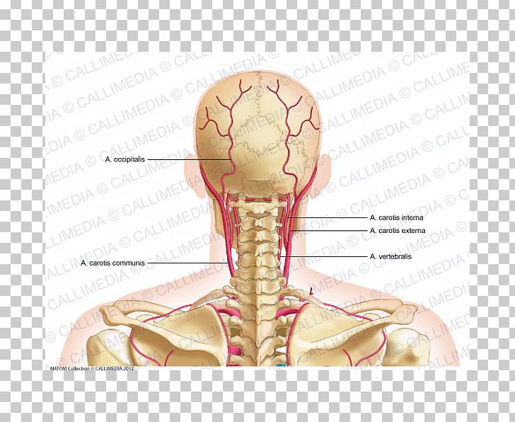
Find this pin and more on anatomy and pysiology block 2by tonna brinson. Superficial dissection of the right side of the neck, showing the carotid and subclavian arteries. This technique involves inserting a catheter into the blocked areas of your carotid arteries. It supplies the thyroid gland. The left and right carotids, and the left and right vertebral arteries. The principal arteries are the carotid and subclavian arteries. Simple labelled illustration depicting the general pathways for the major arteries of the head and neck. The principal arteries are the carotid and subclavian arteries. Transverse cervical artery is labeled, branching from the thyrocervical_trunk. Simple labelled illustration depicting the general pathways for the major arteries of the head and neck. The neck is supplied by arteries other than the carotids. The hypoglossal nerve has been displaced downward in this preparation (lingual artery labeled at center left). Variable in extent, the platysma typically spans the space between the superior margins of pectoralis.
The principal arteries are the carotid and subclavian arteries. The principal arteries are the carotid and subclavian arteries. Always read the nutrition labels in the foods you buy. Left axillary a left vertebral a left carotid sinus right subclavian a. Labels include cephalic vein, brachial artery/vein, basilic vein, musculoskeletal nerve, ulnar collateral artery, radial collateral artery, ulnar nerve/artery/vein the arteries in our body are vessels that flow the blood away from our heart and each one of them is a muscular tube lined by a tissue that is smooth.
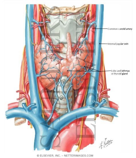
Thyroid Gland and Major Neck Vessels from www.netterimages.com After denudation, all injured carotids of wt mice showed a higher mean. The principal arteries are the carotid and subclavian arteries. The first branch of the thyrocervical trunk is the inferior thyroid artery. The principal arteries are the carotid and subclavian arteries. Arteries of the femoral neck. This technique involves inserting a catheter into the blocked areas of your carotid arteries. In older texts, you might see this referred to as the external the maxillary artery is referred to as the internal maxillary in older textbooks. All veins and arteries are in singular form, this will be easier for the test, i also removed left and right.
Left axillary a left vertebral a left carotid sinus right subclavian a.
After denudation, all injured carotids of wt mice showed a higher mean. Find the perfect neck label stock illustrations from getty images. The principal arteries are the carotid and subclavian arteries. Ninja nerds!join us in this video where we discuss the blood circulation of the head and neck using a flow chart. The easiest spot is where it joins your head, just under the corner of the mandible. Variable in extent, the platysma typically spans the space between the superior margins of pectoralis. The ascending aorta supplies blood to the head, neck, and the arms. We go into great detail on the flow of. Bodytomy provides a labeled celiac artery diagram to help you understand the location, anatomy, and function of this artery. Cholesterol plaque may slowly build up in the carotid artery wall, over decades. Always read the nutrition labels in the foods you buy. Dr calum worsley and assoc prof craig hacking ◉ ◈ et al. Is a delicate, subcutaneous muscle separating the skin from the deeper anterior muscles of the neck.
From this trunk, several vessels arise, which go on to supply the neck. It supplies the thyroid gland. The principal arteries are the carotid and subclavian arteries. The left common carotid artery and left subclavian artery arising directly from the arch of the aorta to supply similar territories on the left side of the body. A blockage in one of the carotid arteries can be cleared either by endarterectomy or carotid angioplasty.
32 Arteries Of The Head And Neck Diagram - Wiring Diagram ... from cdn.imgbin.com Simple labelled illustration depicting the general pathways for the major arteries of the head and neck. From this trunk, several vessels arise, which go on to supply the neck. Depiction of the neck with muscles and arteries shown. Bodytomy provides a labeled celiac artery diagram to help you understand the location, anatomy, and function of this artery. The carotids reside beneath the skin on either side, and the pulse can be felt easily with your hand. In older texts, you might see this referred to as the external the maxillary artery is referred to as the internal maxillary in older textbooks. The easiest spot is where it joins your head, just under the corner of the mandible. Find the perfect neck label stock illustrations from getty images.
Ninja nerds!join us in this video where we discuss the blood circulation of the head and neck using a flow chart.
The carotids reside beneath the skin on either side, and the pulse can be felt easily with your hand. Arteries of the femoral neck. The coronary artery and the circumflex artery are responsible for delivering oxygenated blood to the heart and break it is also the biggest artery in the human body. Left axillary a left vertebral a left carotid sinus right subclavian a. Cholesterol plaque may slowly build up in the carotid artery wall, over decades. The growing plaque may eventually narrow the carotid artery, known as stenosis, and can lead to a stroke. Labeled diagram of the arteries of the head and neck. From this trunk, several vessels arise, which go on to supply the neck. It descends posterolateral to common and internal carotid arteries and gets the. Neck, in land vertebrates, the portion of the body joining the head to the shoulders and chest. Arteries of femoral head … Labels include cephalic vein, brachial artery/vein, basilic vein, musculoskeletal nerve, ulnar collateral artery, radial collateral artery, ulnar nerve/artery/vein the arteries in our body are vessels that flow the blood away from our heart and each one of them is a muscular tube lined by a tissue that is smooth. There are 4 main arteries in your neck;
Ninja nerds!join us in this video where we discuss the blood circulation of the head and neck using a flow chart arteries in neck. Label the arteries of the neck in the ct angiogram.


0 Komentar